Wikimedi'Òc
Modes d'emploi

Cet album fait partie des albums
Cet album photos contient les sous-albums suivants :


Hodenschema.svg - Uwe Gille

Gonadenanlage1.svg - Uwe Gille

Germinal epithelium testicle.svg - Uwe Gille
En-us-testicle.ogg - Vanished user 58234729

Desarrollo Testicular.png - Erikapuentes
Rete testis.jpg - KDS444
Mll5-Is-Required-for-Normal-Spermatogenesis-pone.0027127.s012.ogv - Open Access Media Importer Bot
OAZ-tOAZ3-Is-Essential-for-Rigid-Connection-of-Sperm-Tails-to-Heads-in-Mouse-pgen.1000712.s004.ogv - Open Access Media Importer Bot
Spermatogonial-Stem-Cell-Niche-and-Spermatogonial-Stem-Cell-Transplantation-in-Zebrafish-pone.0012808.s008.ogv - Open Access Media Importer Bot
Spermatogonial-Stem-Cell-Niche-and-Spermatogonial-Stem-Cell-Transplantation-in-Zebrafish-pone.0012808.s009.ogv - Open Access Media Importer Bot
Spermatogonial-Stem-Cell-Niche-and-Spermatogonial-Stem-Cell-Transplantation-in-Zebrafish-pone.0012808.s010.ogv - Open Access Media Importer Bot
Spermatogonial-Stem-Cell-Niche-and-Spermatogonial-Stem-Cell-Transplantation-in-Zebrafish-pone.0012808.s011.ogv - Open Access Media Importer Bot
Spermatogonial-Stem-Cell-Niche-and-Spermatogonial-Stem-Cell-Transplantation-in-Zebrafish-pone.0012808.s012.ogv - Open Access Media Importer Bot
Mammalian-Sperm-Head-Formation-Involves-Different-Polarization-of-Two-Novel-LINC-Complexes-pone.0012072.s005.ogv - Open Access Media Importer Bot
In-vivo-Bioimaging-as-a-Novel-Strategy-to-Detect-Doxorubicin-Induced-Damage-to-Gonadal-Blood-Vessels-pone.0023492.s003.ogv - Open Access Media Importer Bot
In-vivo-Bioimaging-as-a-Novel-Strategy-to-Detect-Doxorubicin-Induced-Damage-to-Gonadal-Blood-Vessels-pone.0023492.s004.ogv - Open Access Media Importer Bot
Cyclical-and-Patch-Like-GDNF-Distribution-along-the-Basal-Surface-of-Sertoli-Cells-in-Mouse-and-pone.0028367.s006.ogv - Open Access Media Importer Bot
Cyclical-and-Patch-Like-GDNF-Distribution-along-the-Basal-Surface-of-Sertoli-Cells-in-Mouse-and-pone.0028367.s007.ogv - Open Access Media Importer Bot
Sequential-Loading-of-Cohesin-Subunits-during-the-First-Meiotic-Prophase-of-Grasshoppers-pgen.0030028.sv001.ogv - Open Access Media Importer Bot
Sequential-Loading-of-Cohesin-Subunits-during-the-First-Meiotic-Prophase-of-Grasshoppers-pgen.0030028.sv002.ogv - Open Access Media Importer Bot
Invasion-of-Wolbachia-into-Anopheles-and-Other-Insect-Germlines-in-an-Ex-vivo-Organ-Culture-System-pone.0036277.s001.ogv - Open Access Media Importer Bot
Invasion-of-Wolbachia-into-Anopheles-and-Other-Insect-Germlines-in-an-Ex-vivo-Organ-Culture-System-pone.0036277.s002.ogv - Open Access Media Importer Bot
F-actin-based-extensions-of-the-head-cyst-cell-adhere-to-the-maturing-spermatids-to-maintain-them-1741-7007-7-19-S2.ogv - Open Access Media Importer Bot
F-actin-based-extensions-of-the-head-cyst-cell-adhere-to-the-maturing-spermatids-to-maintain-them-1741-7007-7-19-S4.ogv - Open Access Media Importer Bot

Internal male reproductive system in Lethocerus patruelis - ZooKeys-319-119-g001.jpeg - Daniel Mietchen

Mixed Germ Cell Tumor of Testis (3260625567).jpg - File Upload Bot (Magnus Manske)

Mixed Germ Cell Tumor of Testis (w ruler) (3261449706).jpg - File Upload Bot (Magnus Manske)
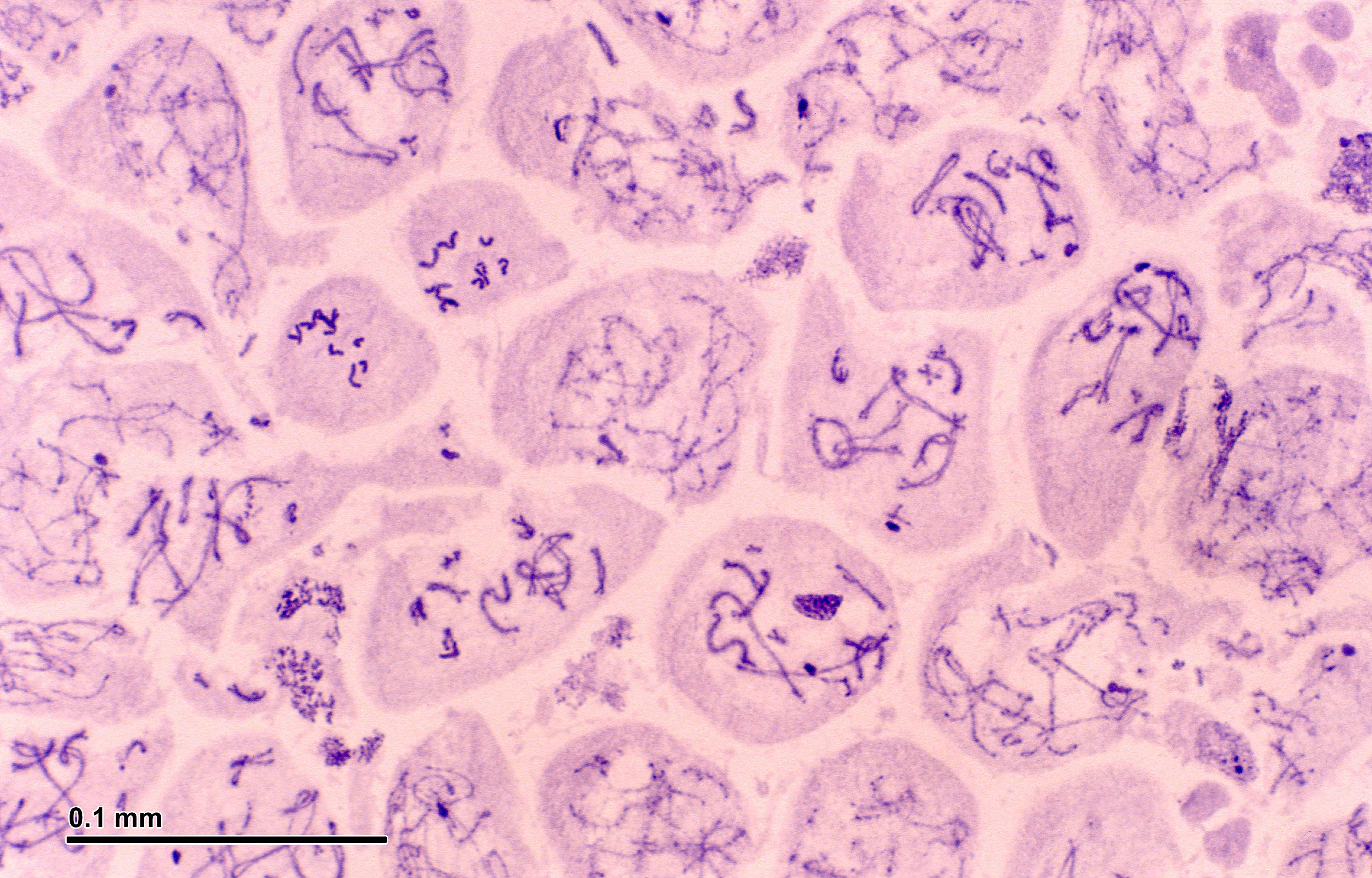
Meiosis (248 23).jpg - Petr Reischig

Essays and observations, physical and literary... Wellcome L0028177.jpg - Fæ

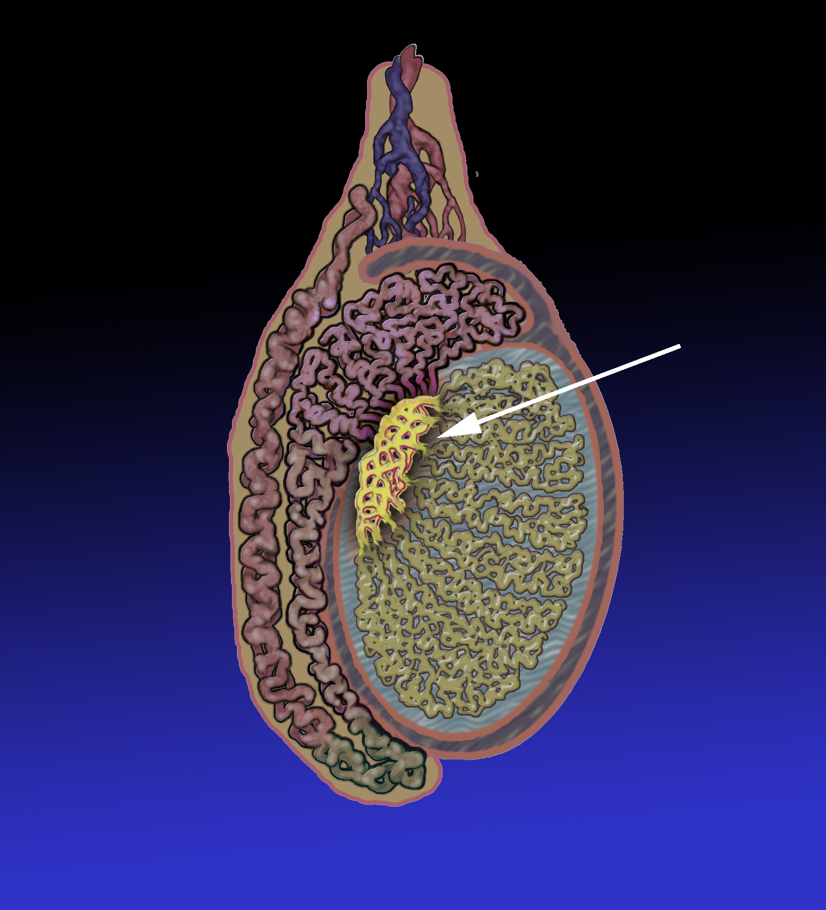
Seminiferous structure ofthe testis and epididymis Wellcome L0040319.jpg - Fæ

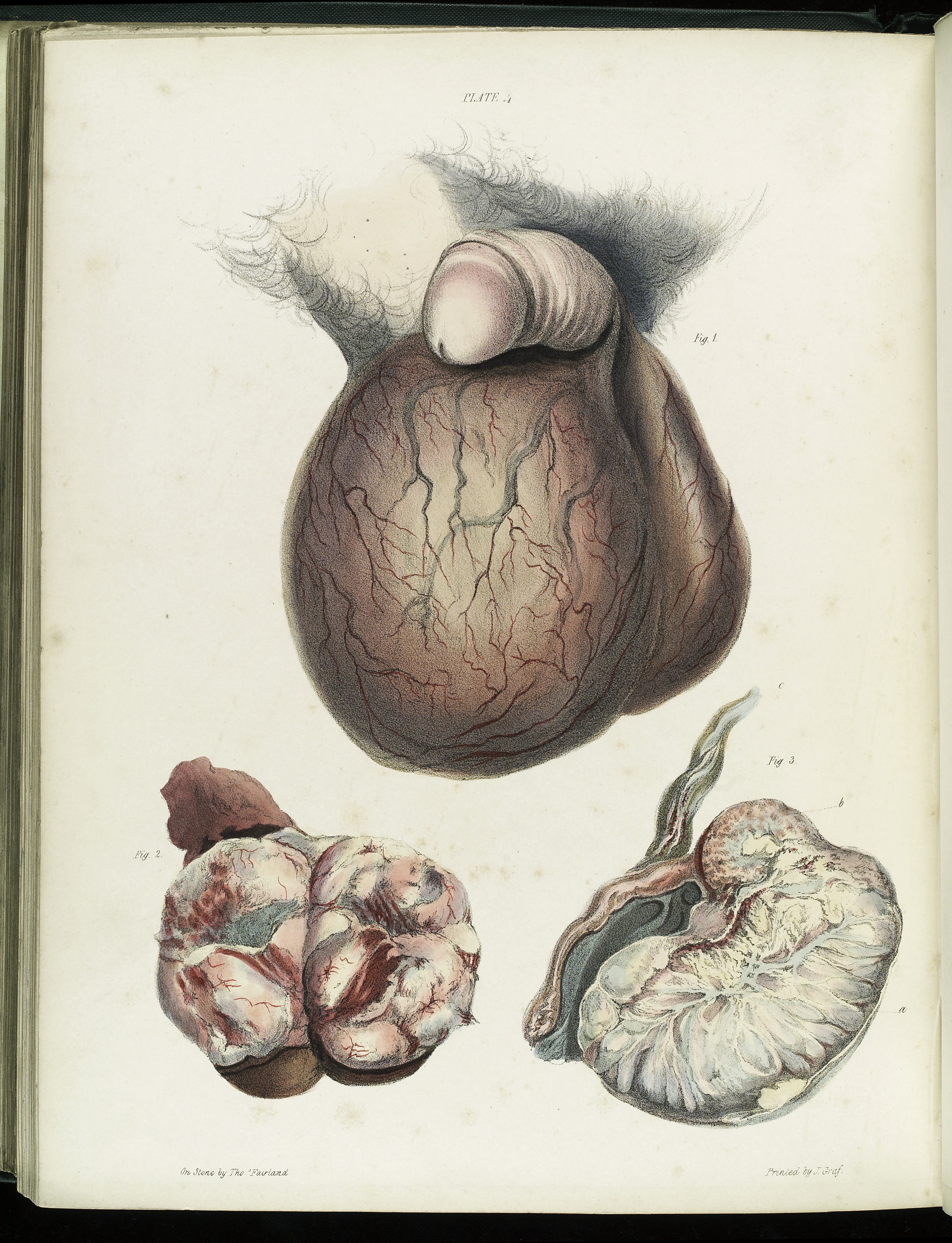
Chronic enlargement of the testis Wellcome L0040320.jpg - Fæ

Inflammation of the testis Wellcome L0040321.jpg - Fæ
Fungoid disease of the testis Wellcome L0040323.jpg - Fæ

Testis with tumours Wellcome L0040324.jpg - Fæ

Illustration of cells infected with Leprosy bacilli Wellcome L0050078.jpg - Fæ

Hoden-Zeichnung.jpg - Volker 71

Lehrbuch der venerischen Krankheiten und der Syphilis (1888) (14784975063).jpg - Fæ

Surgery, its principles and practice (1906) (14771081654).jpg - Fæ

An American text-book of physiology (1897) (14780995834).jpg - Fæ

HHMG - linear.svg - Florent Dufour

Figure 28 01 03.jpg - CFCF

S16-1063 Ranochak, T MGCT- Embryonal + Seminoma + Yolk Sac.jpg - Difu Wu

Image from page 337 of "Chordate morphology" (1962) (19989513674).jpg - Jarble

TDS schemetic diagram.png - Rosieohare
Germinal epithelium slide.jpg - علاء

Primary neuroendocrine tumor of testis.jpg - Netha Hussain

ControlHormFonctionAppGenMasc1.svg - Quo-Fata FERUNT

ControlHormFonctionAppGenMasc2.svg - Quo-Fata FERUNT

Anatomiya gun ku.png - Biyolojiyabikurdi

GFPPupsAndTestisForWiki.jpg - Credd7398

Chickenkidneys.png - Bitran0205

Normal right testis as seen on ultrasound axial view.jpg - Cerevisae

Ultrasound showing left testis calcification.jpg - Cerevisae
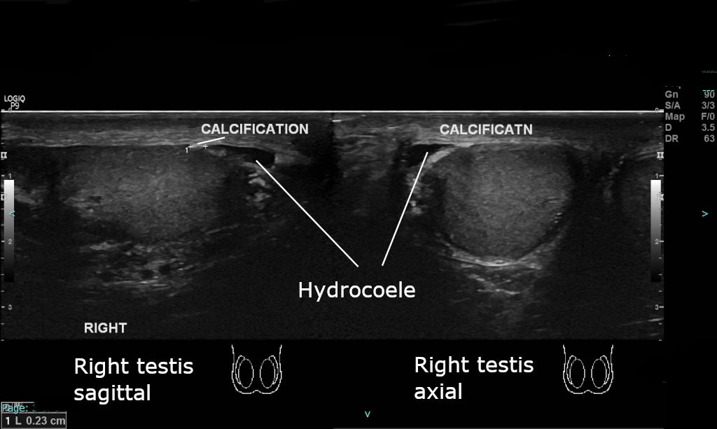
Ultrasound showing right testis calcification.jpg - Cerevisae

Testis eLife.jpg - TiagoLubiana
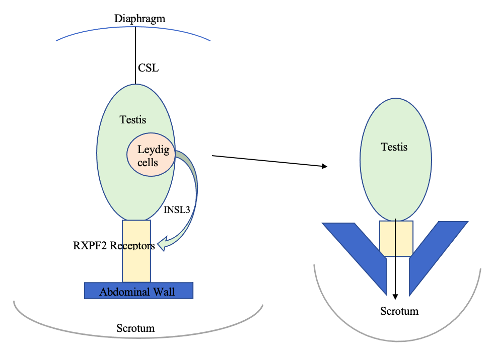
INSL3.Image.png - Sunny1456

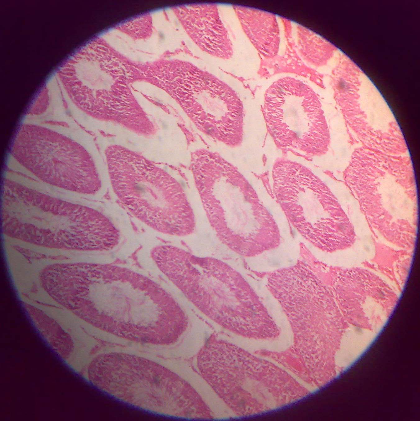
Histology image of testis.jpg - Bhavya akula

MigrazioneTesticolo.png - Rocco Cusari
Transverse section through the left testis and mesonephros of a 20 mm. human embryo. x 250.png - Rasbak

Testicular biopsy shows severely impaired spermatogenesis with an aspect of histological mosaicism. Human.png - Rasbak

Structure and organisation of the human testis and seminiferous tubules.png - Rasbak

3D reconstruction of immunopositive tubules.png - Rasbak

Testis lymphangiogenesis is initiated during late gestation in mice, as visualized by confocal microscopy of whole-mount Prox1-EGFP gonad-mesonephros complexes.png - Rasbak

Lymphatic vessels sprout across, but not beyond, the testis cap at 17.5 dpc.png - Rasbak

Lymphatic vessels are limited to the tunica albuginea in adult testis.png - Rasbak

Lymphatic vessels develop in the postnatal ovary from around 10 dpn.png - Rasbak

The adult ovary possesses an extensive lymphatic network.png - Rasbak

The adult ovary possesses a rich lymphatic network largely overlapping with the blood vasculature.png - Rasbak
Testicular, but not ovarian lymphangiogenesis is initiated during late gestation in mice, as visualized with Prox1-EGFP transgenic gonad-mesonephros complexes.tif - Rasbak
OPT reconstruction showing Prox1-EGFP localization in 17.5 dpc mouse testis as contrasted against ENG-positive blood vessels.ogv - Rasbak
OPT reconstruction showing Prox1-EGFP localization in adult (9-week) mouse ovary contrasted against ENG-positive blood vessels and LYVE1-positive lymphatic capillaries.ogv - Rasbak

Morphology of the human testis. A. Cross section of the human testes.png - Rasbak

Anatomical description of testicular and epididymal structures.jpg - Rasbak
Schematic figure of a transverse section of the seminiferous tubule.jpg - Rasbak

Photomicrograph of the epididymis of the Rhea americana.jpg - Rasbak
Transition of the Rete testis (Rt) and the epididymal ducts (Ep) of the Rhea americana.jpg - Rasbak

Photomicrograph of the Rete testis is formed by cuboidal cells and pseudostratified epithelium of the Rhea americana.jpg - Rasbak

Photomicrograph of the proximal efferent duct exhibiting a simple cuboidal epithelium, with amorphous substance in the cellular surface (Circle) of the Rhea americana.jpg - Rasbak

Epithelium of the distal efferent duct (Ep) of the Rhea americana.jpg - Rasbak

Epithelium of the Rhea americana epididymal duct during the sexual activity period (Ep), which is characterized by pseudo-stratification with the presence or lack of stereocilia.jpg - Rasbak

Photomicrograph of the epididymal ducts of the Rhea americana during sexual repose.jpg - Rasbak

Electrophotomicrograph of elongated spermatids (al) from Rhea Americana, observe the elongation and compacting of the nucleus (arrow).jpg - Rasbak

Architecture of Sertoli cells in the adult mouse seminiferous tubule.jpg - Rasbak

The structure of the mouse testis, the rete testis, and the seminiferous epithelium of the convoluted seminiferous tubule.png - Rasbak

Embryonic development of the mouse rete testis.png - Rasbak
Schematic diagram showing the transitional zone of the convoluted seminiferous tubule and the rete testis from an adult mouse.png - Rasbak

Evaluation of seminiferous tubules in monkey testes using busulfan treatment.png - Rasbak

Ultrasound-guided injection of the rete testis of a monkey.png - Rasbak

Seminiferous tubules are shown in neonatal (6 M), juvenile (18 M), and adult (60 M) rhesus monkey testes.png - Rasbak

Pre-operative ultrasound of present case. Cysts in the right mediastinum testis (black arrow).jpg - Rasbak

Cystic dysplasia of rete testis.jpg - Rasbak

View looking from above into the pelvis and lower part of the abdominal cavity in a fqtus of about the seventh month.jpg - Rasbak

Left testis and epididymis viewed from behind, showing the ductus epididymidis and the first part of the ductus deferens.jpg - Rasbak

Diagram to illustrate the descent of the testis and the manner in which the tunica vaginalis is derived.png - Rasbak

View looking from above into the pelvis and lower part of the abdominal cavity in a foetus of about the seventh month.jpg - Rasbak

Testis. Retroperitoneal mobilisation.png - Rasbak

Fig-09-Amaga pseudobama (Geoplanidae).png - Jeanloujustine

Schematic diagram of spermiogenesis in the testis and epididymis.png - Rasbak