Wikimedi'Òc
Modes d'emploi

Cet album fait partie des albums
Cet album photos contient les sous-albums suivants :


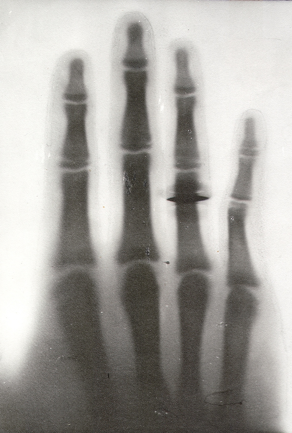
Bound feet (X-ray).jpg - Garrondo

Cardinal vowels-Jones x-ray.jpg - Ish ishwar

WPV X-Ray1.jpg - Doug Coldwell
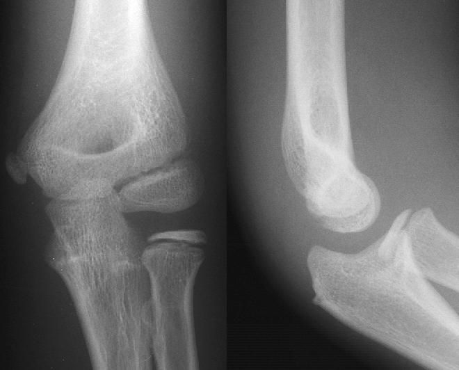
Ap lateral elbow.jpg - File Upload Bot (Magnus Manske)

Vessel-X container.JPG - Aspine

X1300074 Nevit.jpg - Nevit

Medizinische Panoramaaufnahme.jpg - Richard Huber

Ahlbaeck.jpg - File Upload Bot (Magnus Manske)

Gerendertes DVT mit Nervdarstellung.jpg - Dent3D
Gerendertes DVT mit Nervdarstellung klein.jpg - Dent3D

PSM V48 D857 Photo of early x ray.jpg - Ineuw

PSM V50 D678 Effect of 12 second fluorescent screen exposure.jpg - Ineuw

PSM V56 D0681 Dental x ray of a ten year old patient.png - Ineuw

PSM V56 D0682 X ray photo of an elbow joint.png - Ineuw
Pelvis of Albert Fish (X-ray).jpg - Attys

ANAEURISMA0003.jpg - Drmeza

X-rays.jpg - Flickr upload bot

Slide 1.png - Gsjohri

Disc 44.jpg - JonnyD55

World War I radiography amputee.jpg - Hannibal

Hueso trigono II.jpg - Mara Rodriguez Valtierra

Radiograph showing IVC Filter Fracture.jpg - Zackrspv

Gary Simmons' CT scan.jpg - LukeFCartwright

Кишкова непрохідність.JPG - JDiGriz

Кишкова непрохідність..JPG - JDiGriz

Кишкова непрохідність 1.JPG - JDiGriz

Кишкова непрохідність 3.JPG - JDiGriz

Кишкова непрохідність 2.JPG - JDiGriz

Кишкова непрохідність 4.JPG - JDiGriz

Кишкова непрохідність 5.JPG - JDiGriz
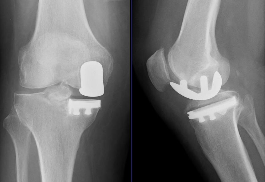
Schlittenprothese.jpg - File Upload Bot (Magnus Manske)
MBq digital-radiograph.jpg - File Upload Bot (Magnus Manske)

3848035360 9a1402df7a bFractureCrâne.jpg - FlickreviewR

The San Pedro Mountain Mummy.jpg - Iamboat

Küntschers Femurnagel von 1940 (1987).JPG - Hellerhoff

Պրոստատի C r-ի ոսկրային մետաստազների արձագանքը կաստրացիայի նկատմամբ.jpg - Doctorzimmer

Radio jambe plus courte.jpg - Dark star

Barium swallow of malignancy oesophagus 02.JPG - Netha Hussain

Barium swallow of malignancy oesophagus 03.JPG - Netha Hussain

Barium swallow of malignancy oesophagus 01.jpg - Netha Hussain

Chest X-Ray.jpg - Zipadyduda
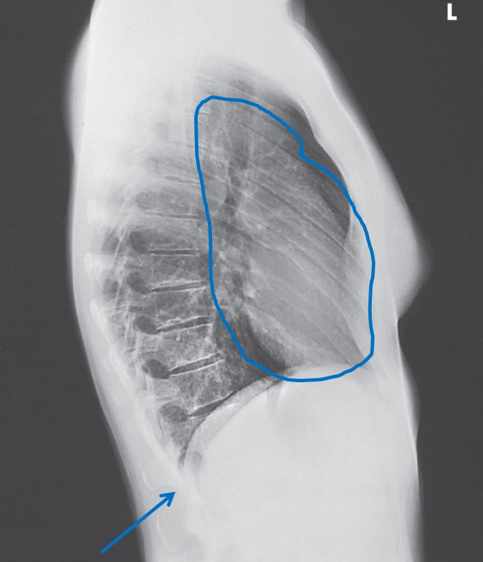
Lateral Chest X-Ray.jpg - Zipadyduda
AP radiograph demonstrating companion shadow of the clavicle.jpg - Jto410
STAR$20Welser.jpg - File Upload Bot (Magnus Manske)

Dislocated Finger XRay.png - Doc James

Typical "Honeycomb lung" x ray- -Cysts appearing like honeycomb, black hyper-inflated lungs and tear drop shaped heart can be seen- (Also there is flattened diaphragm; not visible)- 2014-05-27 04-55.jpg - Phoenix119

X ray internal fixation leg fracture.jpg - Nizil Shah

Hypochondropl 17J W.jpeg - Zieger M

PAVLOVIC MARIJA CR 20130724 90028 1.jpg - VampirehunterD

Перелом со смещением.jpg - Roman2103

The left and right knee-joints of Frank Burgess, probably a Wellcome L0026319.jpg - Fæ

Pelvis of E. Welch, showing disease of right hip and disloca Wellcome L0026321.jpg - Fæ

X-ray of slate pneumoconiosis sufferer's lungs. Wellcome L0029726.jpg - Fæ

C. B. Keetley, Orthopaedic surgery Wellcome L0029866.jpg - Fæ

C. B. Keetley, Orthopaedic surgery Wellcome L0029867.jpg - Fæ

X-Rays, from Handbuch der Geburtshilfe Wellcome L0030715.jpg - Fæ

The bones of the wrist of Mrs Herries; two views. Wellcome L0047930.jpg - Fæ

X-ray of hand deformed by osteo-arthritis. Wellcome M0019124.jpg - Fæ

A woman's pelvis after a pubiotomy - to widen the birth cana Wellcome V0014957.jpg - Fæ

A woman's narrow pelvis with child's head stuck in the middl Wellcome V0014960.jpg - Fæ

X-ray photograph of a skull, probably from a person with Dow Wellcome V0030031.jpg - Fæ

X-ray of an arm, showing a broken bone. Wellcome V0030074.jpg - Fæ
X-ray of a knee joint, showing shrapnel Wellcome V0030078.jpg - Fæ

X-ray of a rib-cage, showing shrapnel Wellcome V0030080.jpg - Fæ

X-ray, skull Wellcome V0030081.jpg - Fæ

X-ray, Alaskan Inuit skull, effects of syphilis, 1910 Wellcome V0031333.jpg - Fæ

מסמר תוך לשדי.jpg - Ofir nahum

X-ray of abdomen with perforated IUD.jpg - Mikael Häggström

Provincial Reconstruction Team, Forward Surgical Team Docs DVIDS246359.jpg - Fæ

Unbehandelter Perthes.jpg - Mehlauge

X-ray3.jpg - OgreBot
Radiography of hand 40 days after hand transplant.jpg - Sonabi
Radiography of the right and left arm after hand transplant.jpg - Sonabi

Anatomischer Anzeiger (1886) (18174086465).jpg - Fæ

X-ray dosage in treatment and radiography (1922) (14758066605).jpg - Fæ

Paragon X-ray pointers - a complete textbook on radiography with Paragon X-ray plates (1915) (14570916187).jpg - Fæ

X-rays simply explained - a handbook on the theory and practice of radiography (1903) (14571415398).jpg - Fæ

X-rays simply explained - a handbook on the theory and practice of radiography (1903) (14755706494).jpg - Fæ

Die Röntgentechnik - Lehrbuch für Ärzte und Studierende (1906) (14571727577).jpg - Fæ

Die Röntgentechnik - Lehrbuch für Ärzte und Studierende (1906) (14755846824).jpg - Fæ

American quarterly of roentgenology (1906) (14777160173).jpg - Fæ

Oral Roentgenology - a Roentgen study of the anatomy and pathology of the oral cavity (1917) (14757821865).jpg - Fæ

Oral Roentgenology - a Roentgen study of the anatomy and pathology of the oral cavity (1917) (14571371407).jpg - Fæ

The itinerary of a breakfast - a popular account of the travels of a breakfast through the food tube and of the ten gates and several stations through which it passes, also of the obstacles which it (14781517964).jpg - Fæ

Interstate medical journal (1917) (14597118678).jpg - Fæ

Roentgen interpretation; a manual for students and practitioners (1919) (14778125713).jpg - Fæ

Interstate medical journal (1917) (14597078289).jpg - Fæ

Interstate medical journal (1917) (14780599001).jpg - Fæ

Interstate medical journal (1917) (14760771296).jpg - Fæ

Interstate medical journal (1917) (14597121738).jpg - Fæ

Interstate medical journal (1917) (14803620633).jpg - Fæ

Interstate medical journal (1917) (14781387534).jpg - Fæ

Interstate medical journal (1917) (14760770846).jpg - Fæ

Interstate medical journal (1917) (14597250157).jpg - Fæ

Journal of radiology (1921) (14571476138).jpg - Fæ

Interpretation of dental and maxillary roentgenograms (1918) (14571564328).jpg - Fæ

Interpretation of dental and maxillary roentgenograms (1918) (14571372020).jpg - Fæ

Interpretation of dental and maxillary roentgenograms (1918) (14754894291).jpg - Fæ

Interstate medical journal (1917) (14597100499).jpg - Fæ

Principles of modern biology (1964) (20744080881).jpg - Fæ

Interstate medical journal (1917) (14783676105).jpg - Fæ

Roentgen interpretation; a manual for students and practitioners (1919) (14754984221).jpg - Fæ

Roentgen interpretation; a manual for students and practitioners (1919) (14571575159).jpg - Fæ

Roentgen interpretation; a manual for students and practitioners (1919) (14571440600).jpg - Fæ

Roentgen interpretation; a manual for students and practitioners (1919) (14571455180).jpg - Fæ

Roentgen interpretation; a manual for students and practitioners (1919) (14571682737).jpg - Fæ

Roentgen interpretation; a manual for students and practitioners (1919) (14571504849).jpg - Fæ

Roentgen interpretation; a manual for students and practitioners (1919) (14757846782).jpg - Fæ

Roentgen interpretation; a manual for students and practitioners (1919) (14777998643).jpg - Fæ

Roentgen interpretation; a manual for students and practitioners (1919) (14571761337).jpg - Fæ

Roentgen interpretation; a manual for students and practitioners (1919) (14778080133).jpg - Fæ

The American annual of photography (1922) (14594948168).jpg - Fæ

Atlas und Grundriss der Unfallheilkunde sowie der Nachkrankheiten der Unfallverletzungen - mit 40 farbigen Tafeln, nach originalquarellen des malers Johann Fink und 141 schwarzen Abbildungen (1900) (14570694618).jpg - Fæ

X-ray observations for foreign bodies and their localisation (1920) (14757764862).jpg - SteinsplitterBot

The Roentgen rays in medicine and surgery as an aid in diagnosis and as a therapeutic agent; designed for the use of practitioners and students (1903) (14758028672).jpg - Fæ

Vegetationsstörungen und Systemerkrankungen der Knochen (1899) (14755801154).jpg - Fæ

The Roentgen rays in medicine and surgery as an aid in diagnosis and as a therapeutic agent - designed for the use of practitioners and students (1901) (14755241344).jpg - Fæ

The Roentgen rays in medicine and surgery as an aid in diagnosis and as a therapeutic agent - designed for the use of practitioners and students (1901) (14734594846).jpg - Fæ

The Roentgen rays in medicine and surgery as an aid in diagnosis and as a therapeutic agent - designed for the use of practitioners and students (1901) (14754450311).jpg - Fæ

The Roentgen rays in medicine and surgery as an aid in diagnosis and as a therapeutic agent - designed for the use of practitioners and students (1901) (14777445863).jpg - Fæ

The Roentgen rays in medicine and surgery as an aid in diagnosis and as a therapeutic agent - designed for the use of practitioners and students (1901) (14757552605).jpg - SteinsplitterBot

The Roentgen rays in medicine and surgery as an aid in diagnosis and as a therapeutic agent - designed for the use of practitioners and students (1901) (14757235232).jpg - SteinsplitterBot

The Roentgen rays in medicine and surgery as an aid in diagnosis and as a therapeutic agent - designed for the use of practitioners and students (1901) (14754411311).jpg - Fæ

The Roentgen rays in medicine and surgery as an aid in diagnosis and as a therapeutic agent - designed for the use of practitioners and students (1901) (14570846270).jpg - Fæ

The Röntgen rays in medical work (1907) (14571065087).jpg - SteinsplitterBot

The Roentgen rays in medicine and surgery as an aid in diagnosis and as a therapeutic agent - designed for the use of practitioners and students (1901) (14570848690).jpg - Fæ

The Roentgen rays in medicine and surgery as an aid in diagnosis and as a therapeutic agent - designed for the use of practitioners and students (1901) (14757506905).jpg - Fæ

The Roentgen rays in medicine and surgery as an aid in diagnosis and as a therapeutic agent - designed for the use of practitioners and students (1901) (14571084797).jpg - Fæ

The Roentgen rays in medicine and surgery as an aid in diagnosis and as a therapeutic agent - designed for the use of practitioners and students (1901) (14570856149).jpg - Fæ

Atlas und Grundriss der Unfallheilkunde sowie der Nachkrankheiten der Unfallverletzungen - mit 40 farbigen Tafeln, nach originalquarellen des malers Johann Fink und 141 schwarzen Abbildungen (1900) (14570647579).jpg - Fæ

The Röntgen rays in medical work (1899) (14733934726).jpg - Fæ

The Röntgen rays in medical work (1899) (14570308648).jpg - Fæ

The Röntgen rays in medical work (1899) (14776809683).jpg - Fæ

The Röntgen rays in medical work (1899) (14733888186).jpg - Fæ

Рентген (перелом ключицы).jpg - DENAMAX

My Back.jpg - CLucy1994

Discograph degenerative disc disease L5-S1.jpg - FastilyClone

Rx de tórax F-Cabello.jpg - SCiardullo
Deacon McGuire hand x-ray.jpg - FastilyClone

Entnahmegebiet im Unterkiefer rechts ohne röntgenologische Auffälligkeit.png - Kipepea

Información de la composición de la imagen.png - Natali Chavez

Afiliado, (sharpening) o filtrado pasa alta.png - Natali Chavez
Transformaciones de intensidad CT.png - Natali Chavez
Radiografía de Luis Zegers - 22 marzo 1896.jpg - ElMachali43
BotonAortico.jpg - SCiardullo

Plain radiograph of the right hip .jpg - FastilyClone

X-ray of Trinket Swallowed by a Child (FDA010) (6946959167).jpg - Vanished Account Byeznhpyxeuztibuo
X- ray with ear ring.jpg - Ibirapuera

Pointmaker Annotation of Digital XRay.jpg - FastilyClone
X-ray of healthy lungs taken at the Institute of Tropical Medicine in Antwerp, on 9 December 2016 (1).tif - Vanished user lkjhgfdsazxcvbnm
X-ray of healthy lungs taken at the Institute of Tropical Medicine in Antwerp, on 9 December 2016 (2).tif - Vanished user lkjhgfdsazxcvbnm
Neumonia.jpg - Maleka Abd Elfatah
Lunatummalazie AP.jpg - Mehlauge

Frauenhand MET DP262472.jpg - Pharos

Hand eines 4 jährigen Kindes MET DP262474.jpg - Pharos

Hand eines 8 jährigen Mädchens MET DP262473.jpg - Pharos

Fuss eines 17 jährigen Jünglings MET DP262475.jpg - Pharos
738a35ed1b3843a28a8be69409b5f6 thumb.jpg - Niteshsah88

111-SC-16187 - NARA - 55192476.jpg - US National Archives bot

Haarverlaengerungen im Roentgenbild - 001.jpg - Hellerhoff

LWS ap.jpg - Couraco

Chest x-ray plain film normal.jpg - Ptrump16

X-Ray of foot joint.jpeg - Deepu.dhaipai

X-ray of Idiopathic interstital pneumonia.jpg - Steve M

Xray of hand showing Ulnar dimelia.jpg - Jim Carter

Knee plain X-ray.jpg - Ptrump16

Cayton.jpg - Haszhasz

Mummy image.jpg - Kulmalukko
Lung X-ray.jpg - Sudraben

XRay R hand print.jpg - Photojack50

XRay R hand with 1st. order equidensities.jpg - Photojack50

AP Whole spine with piercing.jpg - Hollandmjuk

Caput-sourcil angle.jpg - Mikael Häggström

Chest XR of HAPE.png - Maryrosegrant

Esostosi gamba.jpg - AnitaBlakeSZ

Pnemoni-görüntüleme.jpg - Aydinsarihan

Fig2-A-gastrografin-esophagram-shows-a-leak-to-the-left-thoracic-cavity.jpg - Sohmen

بخش بیماران کرونا بیمارستان گلستان نیروی دریایی ارتش 2.jpg - GTVM92

10.1177 0956462414549036-fig7-Secondary yaws- radiographic evidence of osteoperiostitis.jpg - Hellerhoff

Su caso.jpg - Ras67

ProtuberanceEruption 19190529 MeudonSpectroheliograph.jpg - Don-vip

Right Foot of Male human 38 years old side view.png - Gang65

Right Foot of Male human 38 years old top view.png - Gang65
The A B C of the X rays (IA abcofxrays00mead 0).pdf - Fæ

X-Ray Showing Arthrodesis of the Right Wrist.jpg - TheFriendliest

X-Ray on Left Hand Post-Proximal Row Carpectomy 1.jpg - TheFriendliest

X-Ray on Left Hand Post-Proximal Row Carpectomy 2.jpg - TheFriendliest

Left hand of Private John T. Sullivan (CP 3327), National Museum of Health and Medicine (253519649).jpg - Netha Hussain

8822753.fig.001.svg - Steve M
The ABC of the X-rays (IA abcxrays00meadgoog).pdf - Faebot

RX on femur after curettage surgery.jpg - Lucauniverso

RX on femur after curettage surgery 2.jpg - Lucauniverso

פציעה.jpg - Yehud830

Stress-view-of-ankle-with-deltoid-ligament-tear.jpg - Patellaton
Small Lumbar Scoliosis Curve with Pelvic Torque.png - TamrynThrasher

Boy pelvic x-ray pic.jpg - Mahdi2ha

Boy chest x-ray pic.jpg - Mahdi2ha

Chest X-ray of patient with influenza (Reeve 045377), National Museum of Health and Medicine.jpg - Ooligan

X-ray imaging glass plate 24 x 30 cm - probably World War I - Chest torso of a military hospital - Nurse from Germany.jpg - Renamed user f4ce3b16f79cb96621a27f6033316c48

Radiografia-dental.jpg - Rcuellara

Hand X-ray one.jpg - Ser Amantio di Nicolao

Hand X-ray two.jpg - Ser Amantio di Nicolao

Beş aylık cenin, 1896.jpg - Basak

21518 lores.jpg - Ozzie10aaaa

Chest X-ray.jpg - Ptrump16

X-ray General Illustration.jpg - Ptrump16

Radiografía dental panorámica.jpg - Óscar Badillo Pérez

Xray 2022-02-18 15-25-02.png - 1mrln

X-ray of a normal thumb next to a thumb with Brachydactyly type D.jpg - Rollcloud

OtherForComparison.jpg - Doc James

Normal neonatal chest x-ray.jpg - Cerevisae

Chest X-ray showing neurofibromatosis with bilateral pneumonia.jpg - Cerevisae

Ankylosing spondylitis of lumbosacral spine.png - Cerevisae

Chest X-ray showing left lower zone lung mass behind the heart.jpg - Cerevisae

Normal chest radiograph which is under-inspired and is rotated.jpg - Cerevisae

Apparent enlargement of hilum and air space opacities at lung bases due to under inspiration.jpg - Cerevisae

Chest X-ray PA erect showing left upper lobe cavitation.png - Cerevisae

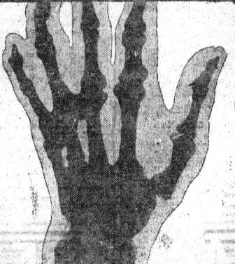
SVG of x-ray of a hand in 1896.svg - Balkanique

Chest X-ray of a normal lung with old left 5th and 6th ribs fractures.jpg - Cerevisae

Cervical x ray.jpg - Challiyan

X ray of Fore arm ap and lat view.jpg - Challiyan

Chest X-ray showing presence of free air under left diaphragm.png - Cerevisae

Fractured Fibula.jpg - Zuluetae24

PMC3202142 CRIM2011-527569.001.png - Ozzie10aaaa

12881 2018 682 Fig1 HTML.png - Ozzie10aaaa

Saltzmann-Aufnahme, junger Erwachsener.jpg - Rasoin
Mesurements Hindfoot Alignment View.jpg - Rasoin

12920 2023 1472 Fig3 HTML.webp - Ozzie10aaaa

X-ray dogs paw ml.jpg - Bahnsen

Congenital lobar emphysema.jpg - Adityagupta95
Congenital cystic adenoid malformation.jpg - Sanjayacuteleukemia
Helsingin yliopiston hammaslääketieteellisen laitoksen röntgenosaston esimies, odontologian tohtori Yrjö Paatero röntgenkuvia tutkimassa 1953 (KK5596-4.HK.8).tif - FinnaUploadBot

Pulmonary Tularemia.png - Ozzie10aaaa

Chlamydia psittaci infection.png - Ozzie10aaaa