Wikimedi'Òc
Modes d'emploi

Cet album fait partie des albums
Cet album photos contient les sous-albums suivants :


NewADHDpic.gif - Fran Rogers
Mozgowie czlowieka mri.png - Neuroscience~commonswiki
Parasagittal MRI of human head in patient with benign familial macrocephaly prior to brain injury (ANIMATED).gif - Dwayne Reed

PET Normal brain.jpg - LeadSongDog
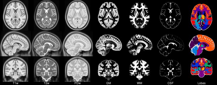
Mni icbm152 sym 09c small.jpg - VladimirFonov

CO2-O2-fMRI-all-over-time.png - Mietchen

Mri head 3dani 1 bionerd.gif - Bionerd

MRI cingulate cortex.png - Jul059
MRI EGC sagittal.png - Codina
BrainMRI3planes.gif - Brainscandude

Connectome.jpg - File Upload Bot (Magnus Manske)
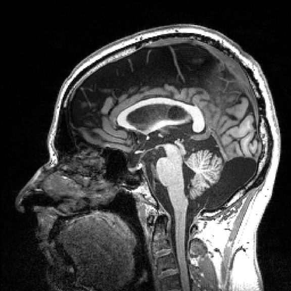
Saghead.jpeg - BiomedNMR
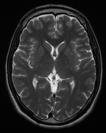
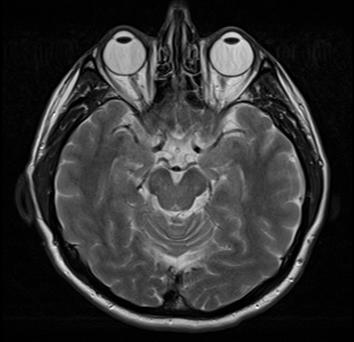
Normal axial T2-weighted MR image of the brain.jpg - Mikael Häggström

Dti-MRI-brain-section.png - Mim.cis
Cabeza del caudado.tif - SCiardullo

Cabeza del caudado.jpg - SCiardullo

IMAGE BRAIN 2.png - Pizzaman1995

Maqnit Rezonans şəkilləri.png - Hüseyn823

Journal.pone .0071275.g002.png - Hüseyn823

Gr1.jpg - Kalona nix

Zona neuretes.jpg - Camendeu

Sinus Habilis Gehirn.jpg - Dr.Komota
Sagittal brain MRI.jpg - TamiresAnsanelo
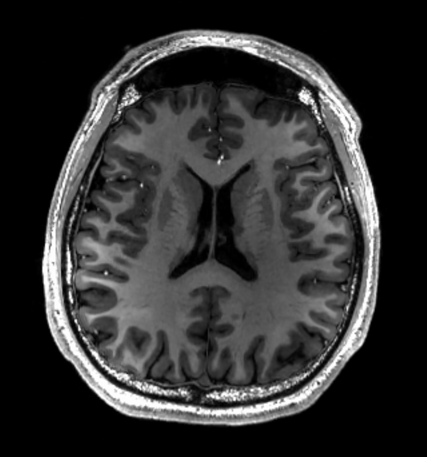
MRI Head Brain Normal.jpg - Ptrump16

MRI brain surface normal.jpg - Ptrump16

Sex in MRI scan.JPG - MARKELLOS
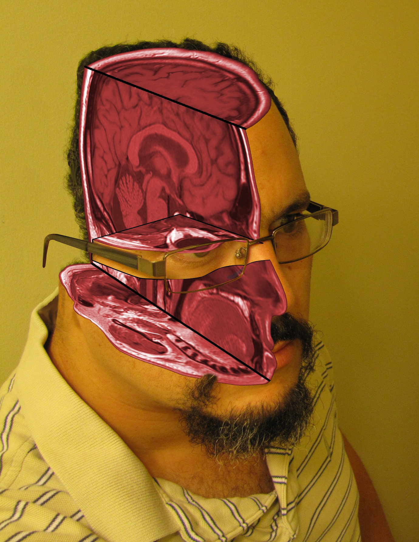
Tomas Diaz MRI self portrait 01.jpg - Tomasdiazdavila
7 Tesla MRI of the ex vivo human brain at 100 micron resolution (100 micron MRI acquired FA25 sagittal) (downsized, original speed).gif - Prototyperspective
7 Tesla MRI of the ex vivo human brain at 100 micron resolution (100 micron MRI acquired FA25 sagittal).webm - Jahobr
7 Tesla MRI of the ex vivo human brain at 100 micron resolution (axial, coronal, sagittal and descriptive summary).webm - Jahobr
7 Tesla MRI of the ex vivo human brain at 100 micron resolution (100 micron MRI acquired FA25 axial).webm - Jahobr
7 Tesla MRI of the ex vivo human brain at 100 micron resolution (100 micron MRI acquired FA25 coronal).webm - Jahobr
Axonal nerve fibers in a brain - the neural network that is us.webm - Prototyperspective

Left Hippocampal Sclerosis on MRI.jpg - Uhomachinky
Angeo2.jpg - 123df

Angeo3.jpg - 123df

Meningitis criptocócica diseminada.png - JOAN
MRI of Human Brain.jpg - Asnaebsa

CT scan and MRI brain image of basal ganglia at CKD and AKI patient.png - LR0725

MRI Brain axial FLAIR sequence showing lentiform fork sign.png - LR0725