Wikimedi'Òc
Modes d'emploi

Cet album fait partie des albums
Cet album photos contient les sous-albums suivants :


Rete testis high mag.jpg - Nephron

Rete testis with seminoma.jpg - Nephron

Adenomatous hyperplasia of the rete testis -- very low mag.jpg - Librepath

Adenomatous hyperplasia of the rete testis -- low mag.jpg - Librepath

Adenomatous hyperplasia of the rete testis -- intermed mag.jpg - Librepath

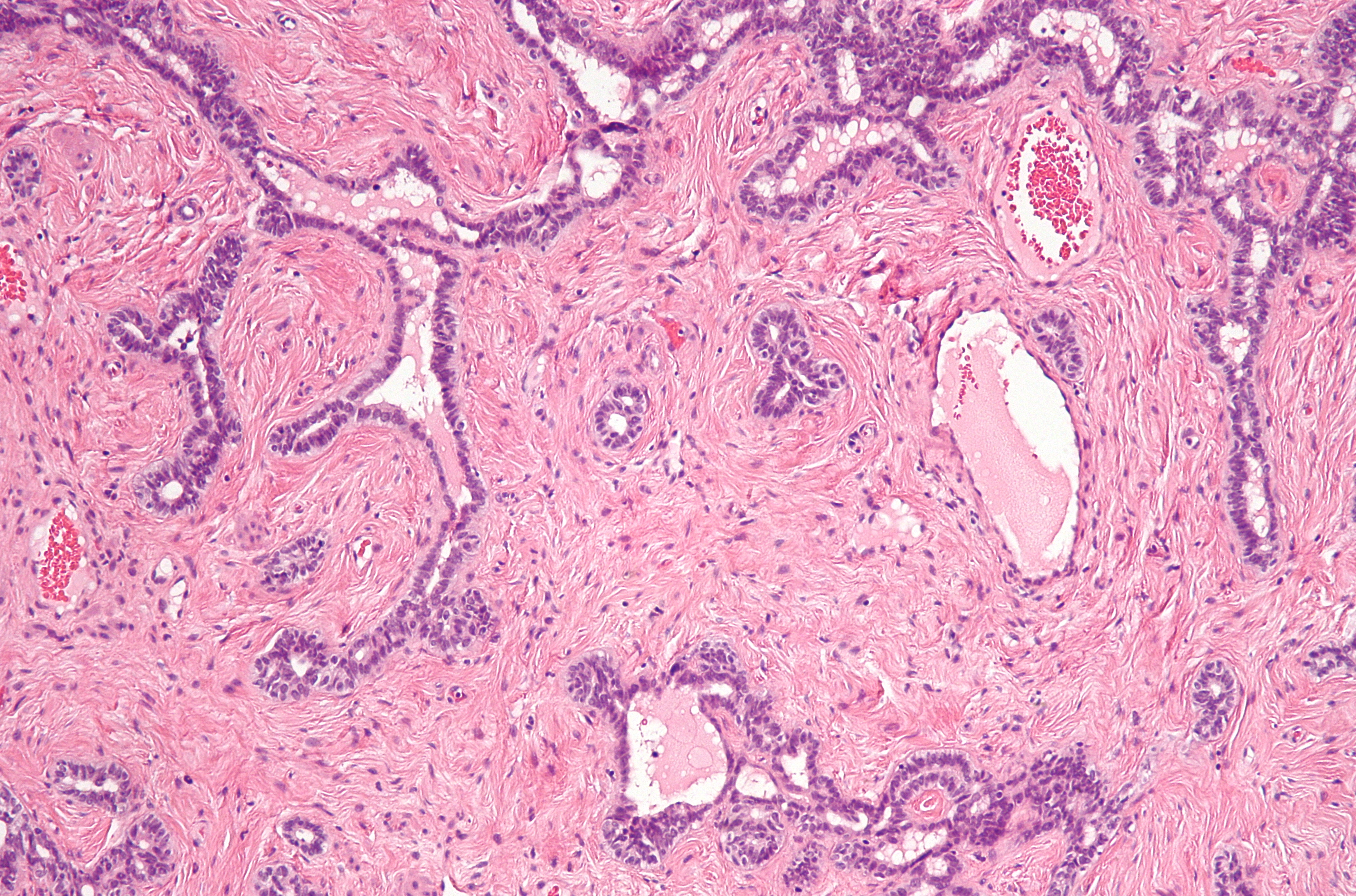
Adenomatous hyperplasia of the rete testis -- high mag.jpg - Librepath

Adenomatous hyperplasia of the rete testis - alt -- low mag.jpg - Librepath
Spermato20x.jpg - Dawnmdmd

Photomicrograph of the epididymis of the Rhea americana.jpg - Rasbak
Transition of the Rete testis (Rt) and the epididymal ducts (Ep) of the Rhea americana.jpg - Rasbak

Photomicrograph of the Rete testis is formed by cuboidal cells and pseudostratified epithelium of the Rhea americana.jpg - Rasbak

Photomicrograph of the proximal efferent duct exhibiting a simple cuboidal epithelium, with amorphous substance in the cellular surface (Circle) of the Rhea americana.jpg - Rasbak

Epithelium of the distal efferent duct (Ep) of the Rhea americana.jpg - Rasbak

Epithelium of the Rhea americana epididymal duct during the sexual activity period (Ep), which is characterized by pseudo-stratification with the presence or lack of stereocilia.jpg - Rasbak

Photomicrograph of the epididymal ducts of the Rhea americana during sexual repose.jpg - Rasbak

The structure of the mouse testis, the rete testis, and the seminiferous epithelium of the convoluted seminiferous tubule.png - Rasbak

Embryonic development of the mouse rete testis.png - Rasbak
Schematic diagram showing the transitional zone of the convoluted seminiferous tubule and the rete testis from an adult mouse.png - Rasbak