Wikimedi'Òc
Modes d'emploi

Cet album fait partie des albums
Cet album photos contient les sous-albums suivants :


The Umbilical Cord.jpg - Tilifa Ocaufa

Umbilical cord histology.jpg - Netha Hussain

Happy Halloween! (284183160).jpg - File Upload Bot (Magnus Manske)

Umbilical cord (254 20).jpg - Petr Reischig

Cross section of the umbilical cord.jpg - Johnlancer123

Rôsolovité väzivo, Wharton's jelly - histológia, histology.jpg - Falty14

Стаз крови в пупочных артериях пупочного канатика, увеличение 40, гематоксилин - эозин.jpg - Андрюша Романов

Tejido conjuntivo embrionario mucoso.jpg - Ettlingert
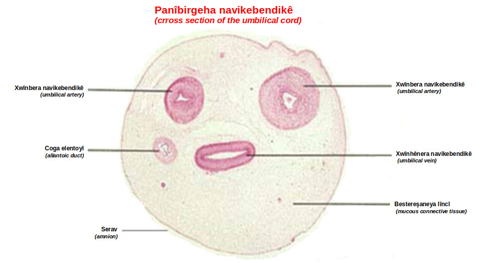
Cross section of the umbilical cord ku.jpg - Biyolojiyabikurdi

Morphology of MSCs derived from WJ, PV, SA, AM and MC in primary culture.png - Rasbak

Histological cross-sections of the human umbilical cord showing the various regions -Wharton’s jelly (WJ), perivascular area (PV), subamnion (SA), amnion (AM)- from which MSCs were derived.png - Rasbak

Cross-sectional diagram of human umbilical cord shows anatomical compartments, including Wharton’s jelly, as a source of stem cells.png - Rasbak

Cross section of human umbilical cord covered with the umbilical cord lining, with an outer layer of umbilical epithelium, and three umbilical vessels embedded in Wharton’s jelly.jpg - Rasbak

Characterization of Decellularized Wharton’s Jelly Matrix (DWJM) A) A fragment of the isolated DWJM.png - Rasbak
Working model of Wharton’s jelly mesenchymal stem cells + PF127 + SAP transplantation in diabetic rats.png - Rasbak

Histology analysis of the dermis regeneration and collagen deposition rat.jpg - Rasbak
Time-lapse imaging of WJMSCs seeded on DWJM. rat.ogv - Rasbak
Time-lapse imaging of WJMSCs on DWJM in different Z planes. rat.ogv - Rasbak

Umbilical cord-derived mesenchym stemcells (MSCs)—location and characteristics.png - Rasbak

Morphological appearance of human amniotic epithelial cells AECs and umbilical cord.jpg - Rasbak