Wikimedi'Òc
Modes d'emploi

Cet album fait partie des albums
Cet album photos contient les sous-albums suivants :


Baihokkhi kakkhi.jpg - Albert~commonswiki

Dentition1.JPG - Ixitixel

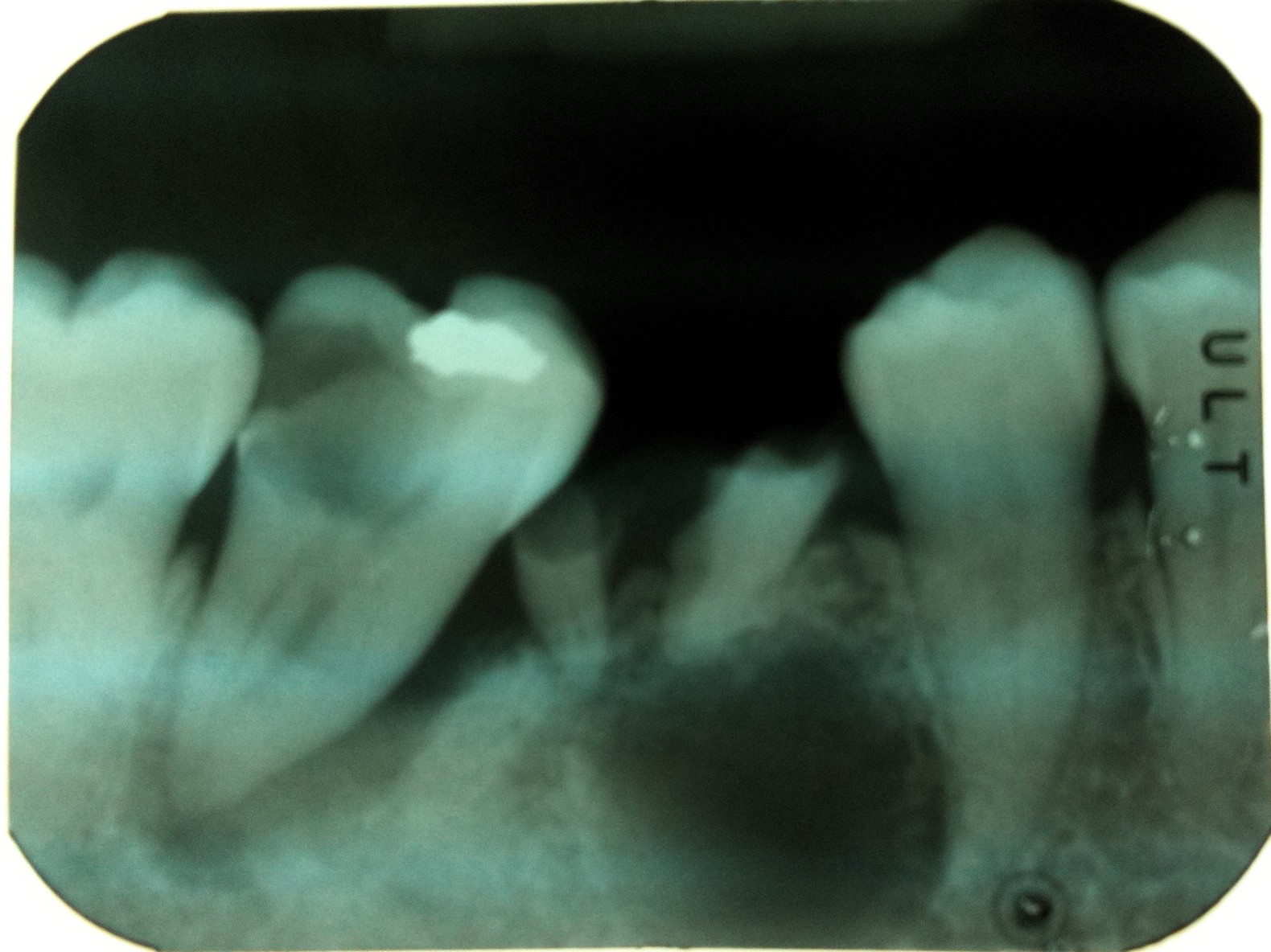
Dental X-ray105.JPG - Werneuchen
Dental X-ray106.JPG - Werneuchen
Dental X-ray108.JPG - Werneuchen

Root resorption.JPG - Dentlavkesh

US Navy 021201-N-5362F-003 Dental Technician inspects a patient's dental x-rays for signs of tooth decay.jpg - BotMultichillT

US Navy 061216-N-5345W-018 General dentistry officer Lt. Adam Clock looks over x-rays while taking care of a patient in the dental lab aboard the Nimitz-class aircraft carrier USS Harry S. Truman (CVN 75).jpg - BotMultichillT

US Navy 040127-N-5134H-001 Using an Intraoral Radiography machine, Dental Technician Ramona Loporto, X-ray's Photographer's Mate 2nd Class Michael Watkins, teeth and gum lines during a routine dental examination.jpg - BotMultichillT
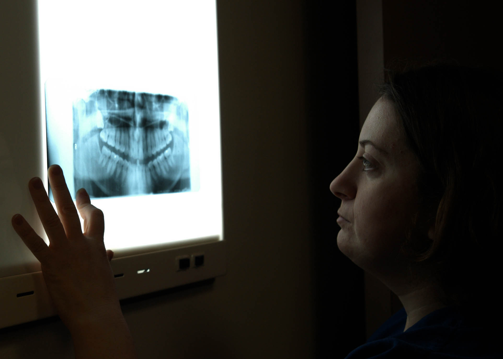
US Navy 040127-N-5134H-002 Dental Technician Ramon Loporto, reviews the X-ray taken of her patient's teeth.jpg - BotMultichillT

Eckzahn retiniert und verlagert Zahn 13 OPG 20100106 001.JPG - Politikaner

Eckzahn retiniert und verlagert Zahn 13 OPG 20100106 003.JPG - Politikaner

Dental imaging UTHSCSA1.JPG - Nightryder84

US Navy 110113-N-6999H-072 Dr. William Reynolds checks a patient's x-rays prior to an examination at Naval Health Clinic Charleston.jpg - BotMultichillT
Zdravé zuby.jpg - Michelina~commonswiki
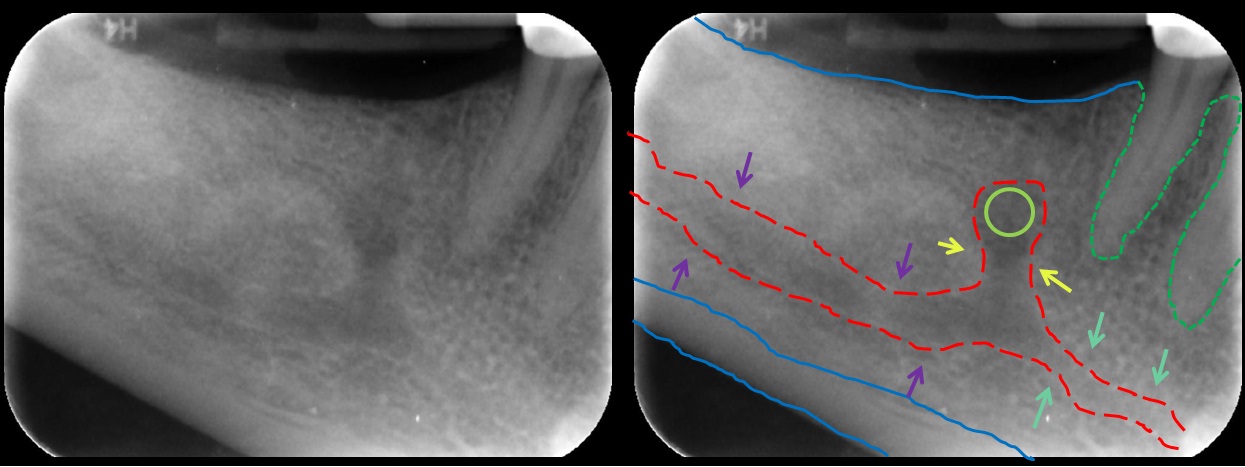
Mandibular Incisive Canal Highlighted.jpg - KMT

US Navy 120131-N-JN664-014 Hospital Corpsman 1st Class Laura Blanco takes X-rays of Electrician's Mate 1st Class Cory Hartley aboard the Nimitz-cla.jpg - BotMultichillT

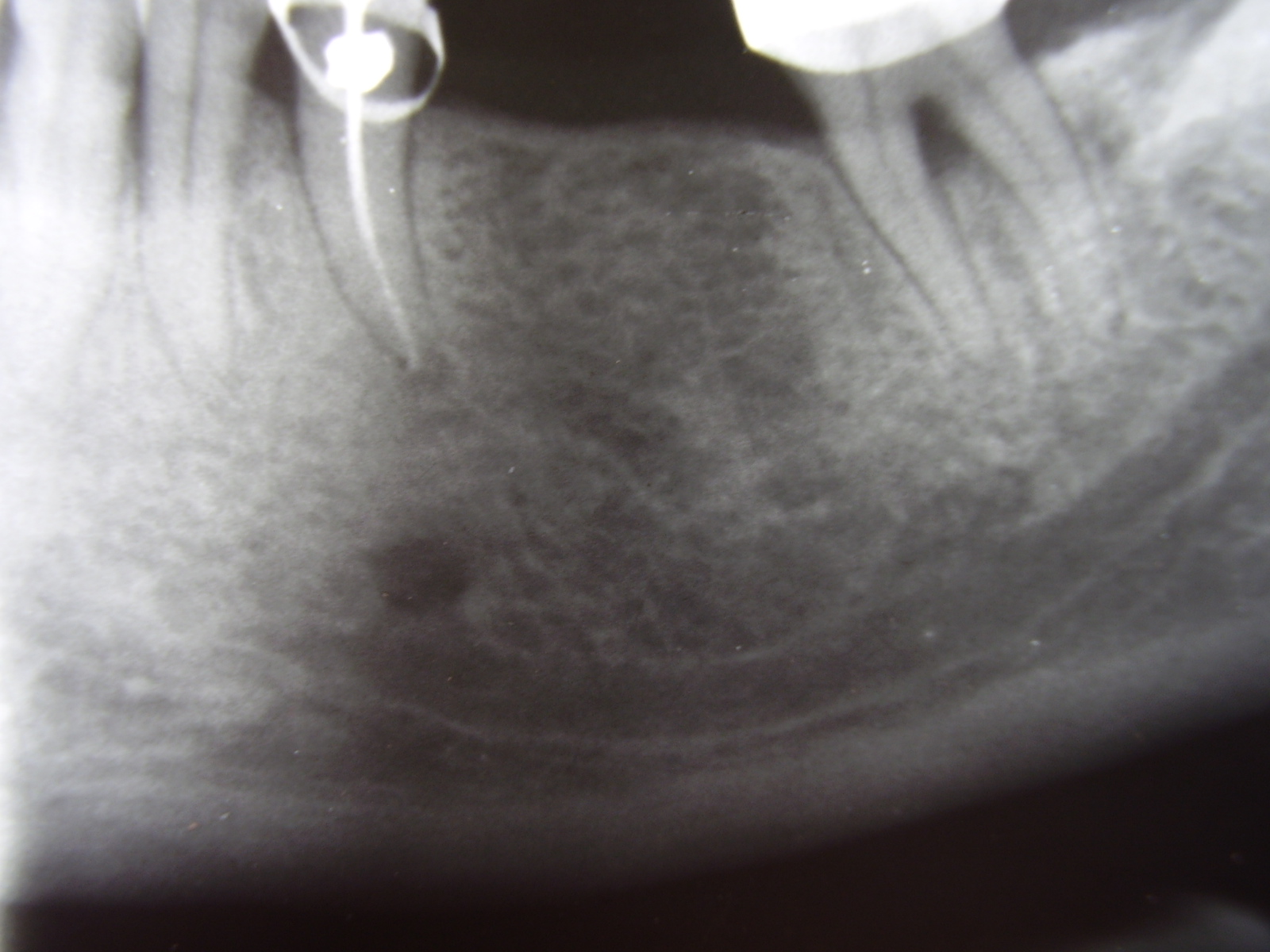
Aufhellung.jpg - File Upload Bot (Magnus Manske)

Chronic apical periodontitis.jpg - Lesion

OPG IMG 6690.jpg - Hellerhoff
Perio-endo lesion.JPG - Lesion

Dental Records (2001).jpg - Fashomecca

7d bild.jpg - FSV

ZELLFAZE MN01 MP03 002.JPG - Zellfaze

ZELLFAZE MN01 MP02 000.JPG - Zellfaze

ZELLFAZE MN01 MP01 011.JPG - Zellfaze

ZELLFAZE MN01 MP04 006.JPG - Zellfaze

ZELLFAZE MN01 MP05 007.JPG - Zellfaze

ZELLFAZE MN01 MP06 013.JPG - Zellfaze

ZELLFAZE MN01 MP08 010.JPG - Zellfaze

ZELLFAZE MN01 MP07 009.JPG - Zellfaze

ZELLFAZE MN01 MP09 003.JPG - Zellfaze

ZELLFAZE MN01 MP11 004.JPG - Zellfaze
ZELLFAZE MN01 MP12 015.JPG - Zellfaze

ZELLFAZE MN01 MP10 012.JPG - Zellfaze

ZELLFAZE MN01 MP14 008.JPG - Zellfaze

ZELLFAZE MN01 MP15 001.JPG - Zellfaze

ZELLFAZE MN01 MP13 014.JPG - Zellfaze

ZELLFAZE MN01 MP16 005.JPG - Zellfaze

Cephalometric radiograph.JPG - ANUG

Ghabara 13.jpg - Ghabara

USS San Antonio action 130714-N-WX580-069.jpg - Fæ
All necessary medical services 130718-A-BG398-002.jpg - Fæ

Biomechanichal preparation of lower second molar incomplete wl.jpg - Dr.mohan m.muthal

Broken endodontic file in mesial root canal.jpg - Dr.mohan m.muthal

ZELLFAZE MN01 MP15 001 Mark.png - Partynia

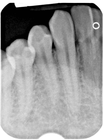
Iopa.jpg - Mmohan7

Apikale Ostitis Zahn 25 2018-05-09.JPG - Bin im Garten

Bone loss in periapical xray.jpg - Shaimaa Abdellatif

Periapical radiolucency.jpg - Shaimaa Abdellatif

X Ray Teeth (PSF).png - Encik Tekateki

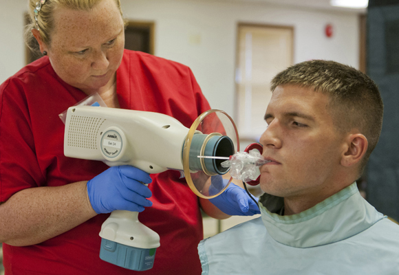
A dental assistant aims an X-ray gun at the mouth of Dental Technician 3rd Class (DT3) Owens in a radiology room in the base dental clinic - DPLA - 9c158451f7e3c29834af028a6e739522.jpeg - DPLA bot

A movable pedestal with "controls" mounted on it.png - Balkanique

Overhead wiring system.png - Balkanique

Radiografía dental panorámica.jpg - Óscar Badillo Pérez

Orthopantomogram of a mixed dentition patient with curved root.jpg - Challiyan

Orthopantomogram of a patient with Eagles syndrome due to impacted upper third molar.jpg - Challiyan

Hemisection of Molar tooth.jpg - Challiyan
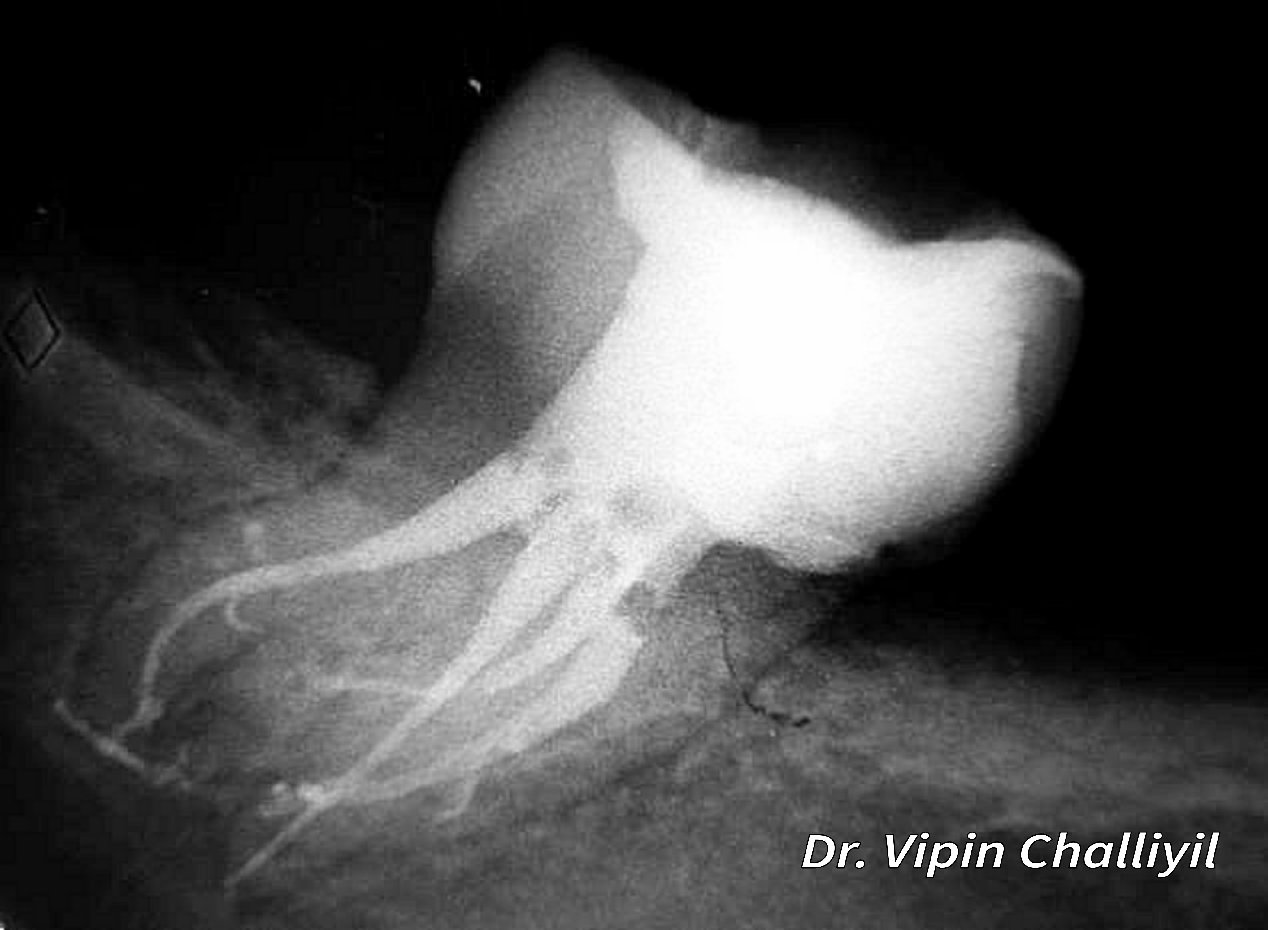
Root canal treatment of lone standing molar.jpg - Challiyan

Teeth, Root canals, Dentistry, Endodontology, Teeth dental X-rays, Rostov-on-Don, Russia.jpg - Argenberg
Impianto.JPG - Finizio

Granuloma sotto dente già devitalizzato - visione di lastra su schermo.jpg - Anna.Massini

Periapical COD.png - Shqynq

Стоматология1.jpg - Aniskov